Diagnostic Investigation
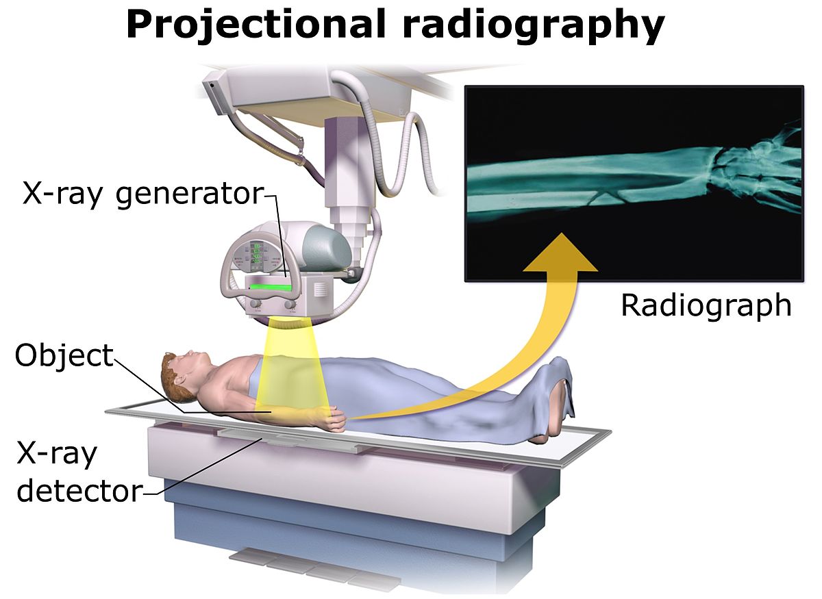
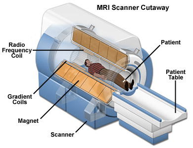
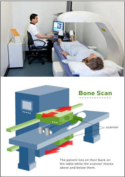
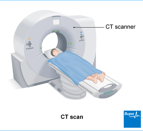
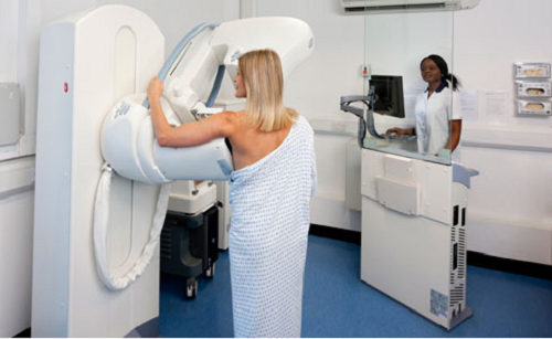
Today, a growing number of tests can be performed to identify the presence of abnormal cells or an abnormal structure. These tests can simply confirm or eliminate a primary cancer (diagnose), or they can help determine the spread of the malignancy (stage). Essentially these investigations fall into three main groups: Radiology, Pathology and Endoscopy.

I. RADIOLOGY

Radiology allows for visualization of the internal structures. Images are created, which the radiologist then interrupts. These images can be created in a number of ways:

X-rays

X-rays or gamma rays are passed through a particular part of the body to generate an image, for example a chest X-ray or mammogram (Yarbro, Frogge and CANCER Goodman, 2005). In order to achieve a clearer image, especially in the gastrointestinal tract, lymphatic vessels, urinary tract and so on, a contrast medium can be used. This involves injecting or, in the case of gastrointestinal studies, asking the patient to swallow a contrast medium. This contrast medium enhances the structures, thereby providing clinicians with more detailed information (Yarbro, Frogge and Goodman, 2005).

Magnetic Resonance Imaging (MRI)

MRI scans use radio waves and strong magnets instead of x-rays. The energy from the radio waves is absorbed and then released in a pattern formed by the type of tissue and by certain diseases. A computer translates the pattern of radio waves given off by the tissues into a very detailed image of parts of the body. A contrast material might be injected just as with CT scans but is used less often. MRI scans are most helpful in looking at the brain. They take longer than CT scans — often up to an hour. The test is painless, but you must lie still inside a cylinder, the surface of which is just a few inches from your face. The machine makes a loud, thumping noise as the magnet switches on and off, so some facilities provide earplugs or headphones to block this sound out.

Nuclear Medicine Imaging

Nuclear medicine imaging involves the parenteral or enteral administration of radioactive compounds. The radioactive material concentrates in the organs or tissues under investigation. A special camera (gamma camera) is used to obtain images of the specific organ/tissues, for interpretation by the radiologist.

Computed Tomography (CT)

The CT scan is an x-ray procedure that produces detailed, cross-sectional images of your body. Instead of taking one picture, as does a conventional x-ray, a CT scanner takes many pictures as it rotates around you. A computer then combines these pictures into an image of a slice of your pelvis. The machine will take pictures of multiple slices of your abdomen. Often, after the first set of pictures is taken, you will receive an intravenous injection of a harmless contrast agent or dye that helps better outline structures in your body. A second set of pictures is then taken. You will need an (intravenous) line through which the contrast dye is injected. The injection can also cause a flushed feeling. Some people are allergic and get hives or, rarely, more serious reactions like trouble breathing and low blood pressure can occur. Be sure to tell the doctor if you have ever had a reaction to any contrast material used for x-rays.

Positron Emission Tomography (PET)

PET scans involve the use of glucose (a form of sugar) that contains a radioactive atom. A small amount of the radioactive material is injected into your arm. Then you are put into the PET machine where a special camera can detect the radioactivity. Because of their high rate of metabolism, cancer cells absorb large amounts of the radioactive sugar. PET is useful when your doctor thinks the cancer has spread, but doesn’t know to where. PET scans can be used instead of several different x-rays because it scans your whole body and may find spread of the cancer where CT scans haven’t. PET scans have become more accurate because newer devices combine the PET scan with a CT scan.

Ultrasound

involves the use of high frequency sound waves. The sound waves are directed over a particular area of the patient’s body, via a probe rubbed over the skin, and echoes are ‘bounced back’. These echoes can be interpreted to provide information relating to the density of the underlying structures. This is particularly useful in distinguishing cysts from more solid structures (Yarbro, Frogge and Goodman, 2005).

II. PATHOLOGY

Pathology tests can confirm a clinical diagnosis, and have been used recently to monitor a patient’s disease and response to treatment. The types of pathology tests that can be undertaken include:

Body fluids such as blood, urine and so on can be used to identify values that fall outside the range expected in a ‘healthy’ individual. For example, an elevated bilirubin and alkaline phosphatase could be indicative of liver disease. Raised calcium could indicate bone metastases.

The production of monoclonal antibodies can lead to the detection of specific tumour antigens, such as HER2 in one type of breast cancer.).

Tumour markers are proteins, antigens, genes or enzymes that can be produced by a tumour. Testing body fluids, including blood, for these markers can be useful in reaching a diagnosis or monitoring an individual’s disease.

When a biopsy is done, tissue is examined directly to see if it has the characteristics of cancer. Tissue is obtained through a needle or a surgical procedure. Biopsy is a good method for diagnosing cancer with certainty.

Cytology involves looking at cells which have been obtained from fluid, secretions, washings from irrigation of cavities, or brushing from tissues.

III) ENDOSCOPY

Endoscopy involves the passage of a long flexible bundle of fibre optic lights. Images are reflected back to the head of the endoscope, providing the operator with a clear picture of the tissues/organs being examined. It is possible for the operator to obtain samples of tissue for histological examination. A pair of special forceps is passed through the endoscope to the area requiring biopsy. The tissue is then retrieved through the endoscope and sent to the laboratory. Cells for cytological examination can also be obtained via this method.